Previous issue | Next issue | Archive
![]() Volume 10 (2S); June 14, 2020 [Booklet] (ICEPF 2020) [EndNote XML for Agris]
Volume 10 (2S); June 14, 2020 [Booklet] (ICEPF 2020) [EndNote XML for Agris]
Assessment of Genetic Variability and Population Structure of Five Rabbit Breeds by Microsatellites Markers Associated with Genes
Rabie TSKM.
J. World Poult. Res. 10(2S): 125-139, 2020; pii: S2322455X2000017-10; DOI: https://dx.doi.org/10.36380/jwpr.2020.17
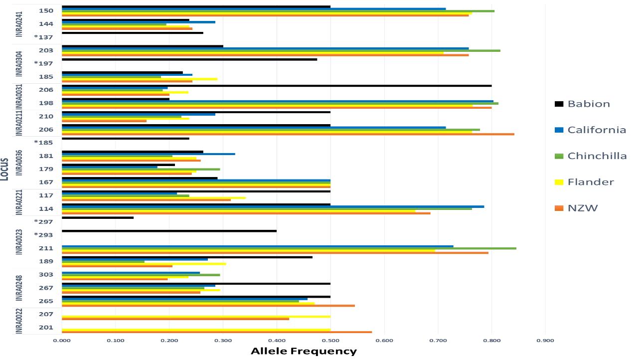
ABSTRACT: The present study was intended to estimate the specific genetic variants by using nine genetic markers among five rabbit breeds (New Zealand White, California, Chinchilla, Flander, and Babion) in Egypt. A total of 128 animals were used (19-35 rabbits per breed). A total of 97 alleles were detected across the breeds and the average number of alleles per locus was 2.16±0.11. Five private alleles were present in Babion breed, where the locus INRACCDDV0023 had two private alleles of 293 and 297 base pairs with allele frequencies of 0.4 and 0.1, respectively. The INRACCDDV0036, INRACCDDV0304, and INRACCDDV0241 loci had private allele for each (185bp (freq: 0.24), 197 (freq: 0.47), and 137bp (freq: 0.26), respectively). The mean of He values ranged from 0.35±0.06 to 0.49±0.07. The average of the polymorphic information content was 0.41 (ranged from 0.298 at INRACCDDV0211 to 0.599 at INRACCDDV0036 locus). To estimate the genetic deviation of the five rabbit breeds, two parameters were evaluated: genetic differentiation (FST), and genetic distance. The FST values varied from 0.029 (INRACCDDV0036) to 0.785 (INRACCDDV0022). The similarity matrix showed that the Chinchilla breed was distinct from other breeds. In addition, among the nine loci, the Hardy-Weinberg equilibrium was highly significant for five loci. Therefore, the rabbit breeds are good reservoirs of allelic diversity that is the major basis for genetic improvement. Consequently, the breeders need a formal conservation plan for such breeds that are in danger of extinction in near future.
Key words: Genetic diversity, Microsatellite marker, Production performance, Rabbits
[Full text-PDF] [XML] [HTML] [ePub] [Crossref Metadata] [Google Scholar]
Mycotoxin Contamination Levels in Broiler Feeds and Aflatoxin Residues in Broiler Tissues
El-Nabarawy AM, Ismael E, Shaaban KhA, El Basuni SS, Batikh MM and Shakal M.
J. World Poult. Res. 10(2S): 133-144, 2020; pii: S2322455X2000018-10; DOI: https://dx.doi.org/10.36380/jwpr.2020.18
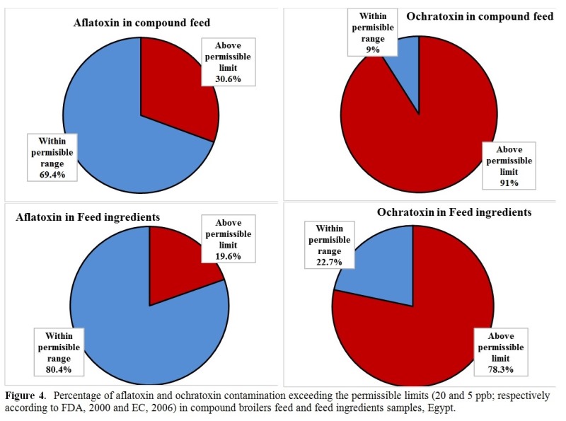
ABSTRACT: The need for regulations to limit the concentration of mycotoxins in feed and food requires the availability of data on levels of contamination in different feedstuffs and estimation of the mycotoxin residues in animal meat. Therefore, this study was conducted to determine contamination levels with different mycotoxins in broiler feed and aflatoxin residues in broilers’ muscle and liver. A total of 194 feed samples, including 148 compound feeds and 46 feed ingredients, were collected from feed manufacturing companies and broiler farms. Feed samples were analyzed for detecting aflatoxins, ochratoxins, zearalenone, and fumonisins using an official analytical method. Moreover, aflatoxin residues were estimated in 64 broiler’s muscle and liver tissues. Obtained results revealed that 100% of compound broiler feed sampled from manufacturing companies were contaminated with aflatoxin and ochratoxin. Also, 96.4% and 92.8% of compound broiler feed sampled from broiler farms were contaminated with aflatoxin and ochratoxin, respectively. Furthermore, 30.6% and 91% of the feed samples were above the permissible levels of aflatoxin and ochratoxin. Aflatoxin residues were detected in all meat and liver samples with levels above the permissible limits. Large scale surveys for determination of different mycotoxins in poultry feed and mycotoxins residues in poultry products are of national and international importance.
Key words: Aflatoxin, Broiler feed, Fumonisin, Mycotoxin residue, Ochratoxins, Zearalenone
[Full text-PDF] [XML] [HTML] [ePub] [Crossref Metadata] [Google Scholar]
Evaluation of Adverse Effects of Antibiotics on Broiler Chickens
Berghiche A, Khenenou T, Bouzid R, Rahem S, Labied I and Boulebda N.
J. World Poult. Res. 10(2S): 145-150, 2020; pii: S2322455X2000019-10; DOI: https://dx.doi.org/10.36380/jwpr.2020.19
ABSTRACT:
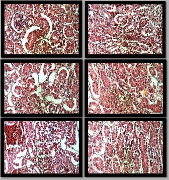
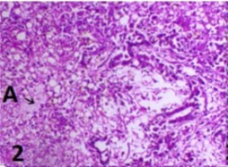
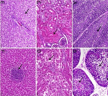
To evaluate the impact of uncontrolled use of veterinary drugs on broilers in eastern Algeria, an experimental plan was developed for the evaluation and identification of drug toxicity in 60 chickens (30 treated and 30 non-treated with antibiotics) using analysis of serum biochemical parameters, autopsy, morpho-metric and histopathological analysis of certain internal organs. The results of the serum biochemical analysis revealed that the uric acid and aspartate aminotransferase values in antibiotic-treated chickens were high, while the lesion status showed a dominance of respiratory lesions, followed by digestive lesions, particularly hepatic lesions. The morphometric study of the internal organs (liver, kidney, and intestine) demonstrated that abnormal liver appearance was very important with minor atrophic changes in the kidney, while the histopathological examination of the liver revealed the presence of deposition in the center of the hexagons in the apical area with an apparent homogeneous structure of fibrous connective tissue. Also, there were apparent deep sinus defects in peripheral areas with an overload of fibrin. The histopathological examination of the kidneys revealed proximal tubular atrophy in the renal parenchyma along with loss of distal intratubular consistency to the peripheral zone of homogeneous structure persuading the peripheral edema. It is concluded that the uncontrolled use of antibiotics in the poultry industry leading to a moderate to severe toxicity.
Key words: Adverse effects, Antibiotics, Broiler chicken, Self-medication
[Full text-PDF] [XML] [HTML] [ePub] [Crossref Metadata] [Google Scholar]
The Effect of Bromhexine and Thyme Oil on Enhancement of the Efficacy of Tilmicosin against Pasteurellosis in Broiler Chickens
Radi AM, Shaban NS, Abo El- Ela FI, Ahmed Mobarez E, El-Gendy AAM and El-Banna HA.
J. World Poult. Res. 10(2S): 151-164, 2020; pii: S2322455X2000020-10; DOI: https://dx.doi.org/10.36380/jwpr.2020.20
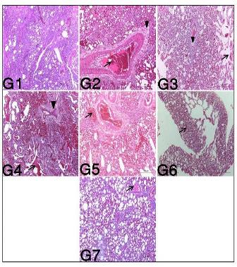
ABSTRACT: Pasteurella multocida is one of the commensal flora of the upper respiratory tract. Under stress conditions, it may be involved as a secondary agent in various respiratory syndromes and caused high mortality as well as significant economic losses in chickens. This study evaluated the effect of bromhexine or thyme oil on enhancement of efficacy of tilmicosin in treatment of avian pasteurellosis. A total of 63 adult chickens were infected by Pasteurella multocida and classified into seven groups and treated as follow; non-infected non-treated group (control negative), infected non-treated group (control positive), group infected and treated by tilmicosin alone, group infected and treated by bromhexine alone, group infected and treated by thyme oil alone, group infected and treated by tilmicosin+bromhexine, and group infected and treated by tilmicosin+thyme oil. Clinical signs, mortality rate, bacterial re-isolation, hematobiochemical and histopathological parameters were determined. The results showed a significant decrease in mortality, bacterial re-isolation as well as clinical signs in combined treated groups compared to tilmicosin group as well as improvement in hematobiochemical and histopathological parameters of combined treated groups. Furthermore, the combination of tilmicosin and bromhexine or thyme oil was more potent in the treatment of pasteurellosis in chickens than each treatment alone. Finally, the clinically observed damage in chickens infected with P. multocida can be ameliorated by a combination of tilmicosin with bromhexine or thyme oil. This protective effect could improve the use of antibiotics in poultry farms as well as reduce human exposure to antibiotic residues and bacterial resistance to antibiotics.
Keywords: Bromhexine, Chickens, Efficacy, Pasteurella Multocida, Thyme oil, Tilmicosin
[Full text-PDF] [XML] [HTML] [ePub] [Crossref Metadata] [Google Scholar]
Antibacterial Sensitivity and Detection of Virulence Associated Gene of Pasteurella multocida Isolated from Rabbits
Mohamed FM, Mansy MF, Abd-Al-Jwad AM and Hassan AK.
J. World Poult. Res. 10(2S): 165-171, 2020; pii: S2322455X2000021-10; DOI: https://dx.doi.org/10.36380/jwpr.2020.21
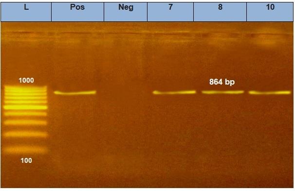
ABSTRACT: The aim of the present work was to determine antibacterial sensitivity and resistance patterns of Pasteurella multocida isolated from rabbits in different farms of Assiut Governorate. Also, this study aimed to detect virulence-associated gene (toxA) of Pasteurella multocida. A total of 40 freshly dead rabbits were used to collect samples from liver, lung and subcutaneous abscess. In addition, tracheal swab samples were collected from 20 diseased rabbits. Bacteriological examination revealed that Pasteurella spp. were isolated and phenotypically identified with an incidence rate of 55% (33 out of 60 rabbits). Ten Pasteurella spp. isolates were randomly chosen for antibiotic sensitivity testing and molecular identification using PCR. Antibiotic sensitivity test was carried using standard disk diffusion method against 13 antibacterial drugs to determine antibacterial sensitivity and resistance patterns of Pasteurella isolates and revealed variable sensitivity and resistance to antibacterial drugs. Pasteurella multocida isolates were sensitive to wide variety of antibiotics (norfloxacin, enrofloxacin, ciprofloxacin, florfenicol, doxycycline, gentamycin, cephradine and cefoxitin). Three out of ten isolates were molecularly confirmed to be Pasteurella multocida and all of them demonstrated the presence of toxA virulence genes. In conclusion, the prevalence of Pasteurella infections in rabbits in Assiut Governorate was relatively high.
Key words: Antibacterial resístanse Pasteurella multocida, toxA gene, virulence genes
[Full text-PDF] [XML] [HTML] [ePub] [Crossref Metadata] [Google Scholar]
The Impact of Alpha-lipoic Acid Dietary Supplementation on Growth Performance, Liver and Bone Efficiency, and Expression Levels of Growth-Regulating Genes in Commercial Broilers
Sakr OA , Nassef E, Fadl SE , Omar H, Waded E , and El-Kassas S.
J. World Poult. Res. 10(2S): 172-179, 2020; pii: S2322455X2000022-10; DOI: https://dx.doi.org/10.36380/jwpr.2020.22
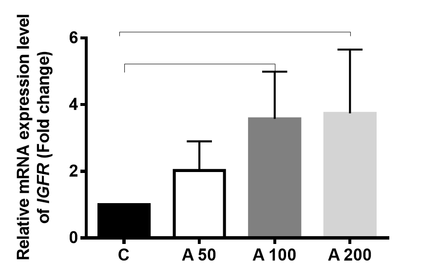
ABSTRACT: Increasing bird growth is a crucial demand for all poultry producers. This occurs by the genetic improvement of the existing breeds and by improving the feeding management. The present study investigated the impact of Alpha-Lipoic Acid (ALA) supplementation in the diet on performance, serum parameters, tibia bone composition, and the expression levels of growth-related genes in chickens. A total of 120 day-old broiler chicks (Cobb 505) were used and divided into four groups. The control group was fed on a basal diet without the ALA supplement. The birds in groups of A50, A100, and A200 were fed on the formulated diet supplemented with ALA at doses of 50, 100, and 200 mg/kg of diet, respectively for 35 days. Results indicated that ALA supplementation significantly improved the birds’ growth performance. This effect was associated with a marked upregulation of mRNA levels of GHR and IGFR and a significant downregulation of MSTN expression level. In addition, the ALA dietary provision caused a distinct improvement in liver function and bone efficiency. Thus, the improving effect of ALA on birds’ growth performance is mediated by modulating the growth-regulating genes. In conclusion, ALA could be used as a good growth-promoter in dietary supplements.
Keywords: Alpha-lipoic Acid; Bone Efficiency; Broilers; Gene Expression; Growth Performance
[Full text-PDF] [XML] [HTML] [ePub] [Crossref Metadata] [Google Scholar]
The Effect of Methionine on Performance, Carcass Characteristics and Gut Morphology of Finisher Broilers under Tropical Environment Conditions
Abdulla NR, Alshelmani MI, Loh TCh, Foo HL and Zainudin MA.
J. World Poult. Res. 10(2S): 180-183, 2020; pii: S2322455X2000023-10; DOI: https://dx.doi.org/10.36380/jwpr.2020.23
ABSTRACT: The present study was conducted to determine the effect of DL- and L-methionine on growth performance, carcass characteristics, and gut morphology during the finisher phase in the tropical environment. A total of 560 one-day-old broiler chicks (Cobb 500) were purchased and raised for 35 days. The chicks were divided into four dietary treatments with seven replicates (20 birds per replicate). The basal diet was offered to the chickens during the starter and finisher phases. The DL-methionine was supplemented to the finisher diet as at 0.260% (T1) and 0.179% (T2). Correspondingly, the L-methionine was supplemented to the finisher diet with the same ratios; 0.260% (T3) and 0.179% (T4). The findings revealed no significant differences in growth performance between the two forms of methionine. The obtained results indicated no significant differences in carcass characteristics, the villi heights and crypt depth among the dietary treatments. In conclusion, DL-methionine can be used in broiler nutrition as substitute for L- methionine which is more expensive in poultry industry.
Key words: Carcass characteristics, Growth performance, Gut morphology, Methionine, Tropical environment
[Full text-PDF] [XML] [HTML] [ePub] [Crossref Metadata] [Google Scholar]
Comparative Clinicopathological Study of Salmonellosis in Integrated Fish-Duck Farming
El-nabarawy AM, Shakal MA, Hegazy AM and Batikh MM.
J. World Poult. Res. 10(2S): 184-194, 2020; pii: S2322455X2000024-10; DOI: https://dx.doi.org/10.36380/jwpr.2020.24
ABSTRACT: Poultry litter is used in fish farms as fertilizer thus integrated fish-duck farming is common in some areas of Egypt. Salmonella bacteria may be present in poultry litter and contaminate fish ponds and infect duck farms. To investigate incidence and prevalence of Salmonella infection in integrated duck-fish farms, 50 litter samples, 200 cloacal swabs from integrated duck farms, 60 liver samples from integrated duck farms and 69 water samples from the fish pond were collected. Results revealed the isolation and identification of 19 Salmonella spp. belonging to 14 different serotypes (4 isolates from litter, 2 isolates from fish pond water, 8 isolates from cloacal swabs of ducks and 5 isolates from ducks liver). Fifty, one-day-old Pekin ducks were experimentally infected with five chosen Salmonella serotypes (S. Bargny, S. Tshingwe, S. Uganda, S. Kentucky, and S. Enteritidis). The results from experimental infection revealed clinicopathological findings including degeneration and necrosis in the liver, lymphoid depletion and macrophage infiltration in the spleen and enteritis. Mortality ranged from 28.6% in S. Bargny, S. Enteritidis and S. Kentucky and increased to 42.9% in S. Uganda and reached up to 100% in S.Tshingwe. Body weight gain decreased by 16% in S. Uganda and exceeded to 23.9% in S. Kentucky and decreased by 31% in S. Bargny and S. Enteritidis as compared to the control group. Feed conversion ratio was recorded and ranged from 5.1, 5.11, 4.98, 5.15 and 4.02 in S. Bargny, S. Uganda, S. Kentucky, S. Enteritidis, and control group, respectively. In conclusion, different species of Salmonella can affect integrated duck-fish farms and cause high mortality as well as a decrease in feed intake, feed conversion ratio, and body weight gain.
Key words: Histopathology, Integrated duck-fish farms, Pathogenicity, Salmonella spp.
[Full text-PDF] [XML] [HTML] [ePub] [Crossref Metadata] [Google Scholar]
Molecular Identification of a velogenic Newcastle Disease Virus Strain Isolated from Egypt
Shakal M, Maher M, Metwally AS, AbdelSabour MA, Madbbouly YM, Safwat G.
J. World Poult. Res. 10(2S): 195-202, 2020; pii: S2322455X2000025-10; DOI: https://dx.doi.org/10.36380/jwpr.2020.25
ABSTRACT: Newcastle Disease Virus (NDV) is still a major concern for the Egyptian poultry industry in spite of the mass vaccination programs implemented from a long years ago. The current study aimed to carry out the molecular identification of surface glycoprotein genes of NDV field strain isolated from the Giza governorate, Egypt. Tracheae were collected from 10 broilers NDV-vaccinated chicken flocks (at least three samples from each flock) suffering from mild to moderate respiratory symptoms; with mortalities varying from 10-40% during October 2019. Only five samples showed HA positive activity after propagation in specific pathogen-free embryonated chicken eggs and only one sample was positive for Avian avulavirus 1 by real-time reverse transcription-PCR. Sequencing for the cleavage site of the F protein gene of the positive isolate showed the typical known sequence of velogenic NDV strains (112RRQKRF117). Phylogenetic analysis of both F and HN genes showed high similarity and close relation to Chinese strains of Genotype VII and more specifically subtype VIId, suggesting the role of migratory wild birds in NDV evolution in Egypt. In conclusion, further epidemiological and surveillance studies are strongly recommended to define the exact role of migratory wild birds in NDV evolution in Egypt.
Key words: Broilers, Newcastle Disease, Poultry industry, Velogenic
[Full text-PDF] [XML] [HTML] [ePub] [Crossref Metadata] [Google Scholar]
Efficacy of Staphylococcus aureus Vaccine in Chicken
El-Maghraby AS, azizSh and Mwafy A.
J. World Poult. Res. 10(2S): 203-213, 2020; pii: S2322455X2000026-10; DOI: https://dx.doi.org/10.36380/jwpr.2020.26
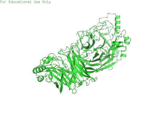
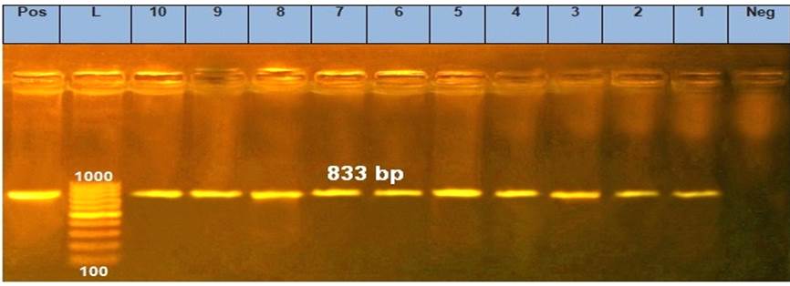
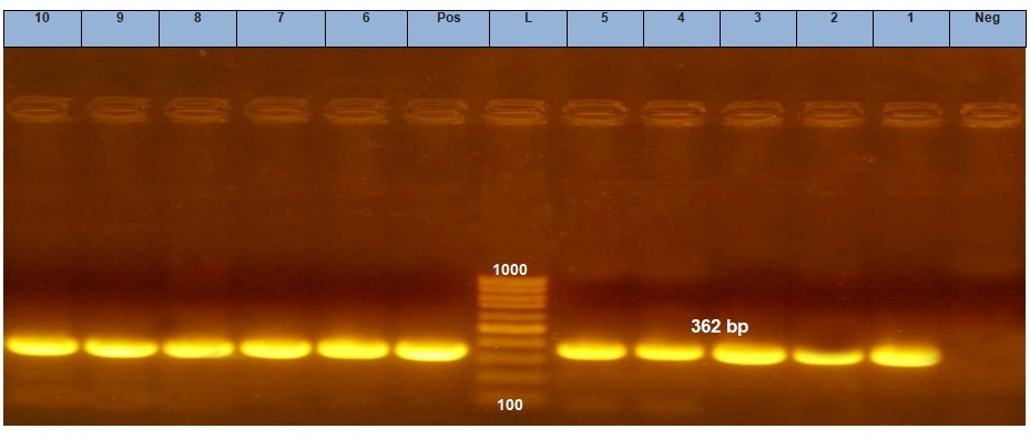
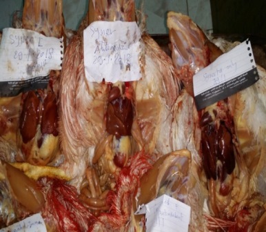
ABSTRACT: Staphylococcus aureus is considered one of the most important pathogens causing septic arthritis in poultry with significant economic losses. This study aimed to evaluate the efficacy of a locally prepared S. aureus vaccine against staphylococcal arthritis in poultry. Out of 78 samples collected from infected chickens showing clinical signs bumble foot, 10 field isolates were detected and confirmed phenotypically by culturing, Gram staining, biochemical and molecular identification to be S. aureus in prevalence of 12.82%. Molecular identification of clumping factor A (ClfA) and blaZ genes of S. aureus isolates revealed that the PCR amplification with ClfA and blaZspecific primers conducted with genomic DNA resulted in products of approximate size 638 bp and 833 bp, respectively. Phylogenetic tree for S. aureus ClfA virulence gene partial sequences was generated using maximum likelihood, neighbour joining and maximum parsimony in MEGA6. It showed clear clustring of Egyptian isolated strain (S. aureus ASM strain) and different S. aureus strains uploaded from GenBank. Sequence identities between the Egyptian isolated strain (S. aureus ASM strain)and different S. aureus strains uploaded from GenBank revealed 99.5% to 100% homology. Also, there was identity and homology in S. aureusblaZgene nucleotide sequence in the Egyptian isolated strain (S. aureus ASM strain)and the different S. aureus strains uploaded from GenBank revealed 96.1% to 98.9% homology. Phylogenetic tree for S. aureusblaZβ-lactamases resistant gene partial sequences showed clear clustring of the Egyptian isolated strain (S. aureus ASM strain)and different S. aureus strains uploaded from GenBank. The results of humoral immune response revealed that the geometric mean antibody values against locally prepared S. aureus vaccine measured by indirect hemagglutination test increased from 1st week post vaccination gradually till reached maximum level (322.5) at 6th week post boostering. The results showed an increased humoral antibody production in vaccinated group that was capable of preventing establishment of new S. aureus infection in vaccinated group compared to control group. The mortality rates in unvaccinated group was higher than that of vaccinated group were (42.5%, vs. 7.5%) at 1st and 2nd week post challenge (39.1% vs. 5.4%). The protection % in challenge assay of the prepared S. aureus vaccine was (92.5% and 87.5%) at 1st and 2nd week post challenge respectively. It could be concluded that the prepeared vaccine was safe, potent and protect birds against S. aureus infection.
Key words: Blaz, ClfA, PCR, Sequencing, Staphylococcus aureus, Vaccine
[Full text-PDF] [XML] [HTML] [ePub] [Crossref Metadata] [Google Scholar]
Association of Antiseptic Resistance Gene (qacEΔ1) with Class 1 Integrons in Salmonella Isolated from Broiler Chickens
Ali NM and Mohamed FM.
J. World Poult. Res. 10(2S): 214-222, 2020; pii: S2322455X2000027-10; DOI: https://dx.doi.org/10.36380/jwpr.2020.27
ABSTRACT: Salmonella enterica is considered a zoonotic pathogen that acquires antibiotic resistance in livestock. In the current study, a total of 18 Salmonella enterica isolates recovered from cloacal swabs of diseased and freshly dead broilers were serotyped and assessed for susceptibility to clinically important antibiotics. The multi-resistant isolates were examined for the presence of the antiseptic resistance genes including quaternary ammonium (qacEΔ1) and class 1 integron-integrase (intI1) by PCR. The results of serotyping of 18 Salmonella isolates indicated that five isolates belonged to Salmonella Typhimurium, four isolates belonged to each of Salmonella Kentucky and Salmonella Enteritidis, three isolates belonged to Salmonella Molade and one isolate belonged to each of Salmonella Inganda and Salmonella Larochelle. Fifteen Salmonella isolates (83.3%) were multi-resistant to at least three antibiotics with a multidrug resistance index value of 0.473. All of the intI1-positive strains carried qacEΔ1, confirming that the qacEΔ1 gene is linked to the integrons. The study concluded that the presence of the qacEΔ1 resistance gene and class 1 integrase in multi-drug resistant Salmonella strains might be contributed to co-resistance or cross-resistance mechanisms.
Key words: intI1, Multidrug-resistant Salmonella, PCR, qacEΔ1
[Full text-PDF] [XML] [HTML] [ePub] [Crossref Metadata] [Google Scholar]
Comparative Evaluation of Different Antimycotoxins for Controlling Mycotoxicosis in Broiler Chickens
El Nabarawy AM , Madian K, Shaheed IB and Abd El-Ghany WA.
J. World Poult. Res. 10(2S): 223-234, 2020; pii: S2322455X2000028-10; DOI: https://dx.doi.org/10.36380/jwpr.2020.28
ABSTRACT:
Natural contamination of feedstuffs with mycotoxins is considered a major problem affecting the poultry industry in Egypt. Accordingly, this study aimed to compare the ability of different antimycotoxin compounds in the control of mycotoxicosis caused by naturally contaminated diet in broiler chickens. A total of 180 day-old broiler chicks were divided into six groups (30 chicks each group) and kept for a 5-week experimental period. Group 1 was kept as control negative (non mycotoxicated or treated), while group 2 was kept as a positive control (mycotoxicated only). Groups 2-6 were fed ration contaminated with 11 ppb aflatoxins, 3.9 ppb ochratoxins, and 4.2 ppm zearalenone. Groups 3-6 were kept in mycotoxicated ration until 2 weeks of age when clinical signs and lesions were suggestive for mycotoxicosis. Groups 3, 4 and 5 were treated with biological, antioxidant, immunostimulant compounds; respectively. Biological, antioxidants and immunostimulant compounds were given in the drinking water. In group 6, ration was treated with formaldehyde vapor. Performance parameters including body weight, feed consumption and feed conversion rate were recorded weekly. Clinical signs, mortalities and lesions were observed. Serum samples were collected for determination of immunological profile to infectious bursal disease (IBD) virus vaccine. Moreover, liver, kidney and bursa of Fabricius were collected for histopathological examination. Muscles and liver tissue samples were collected for determination of aflatoxins residues. Results revealed significant improvement in performance parameters in treated groups in comparison to non-treated mycotoxicated group, however, antioxidants-treated birds showed the highest performance. The severity of clinical signs and lesions were reduced in the treated chickens compared to non-treated mycotoxicated ones. Significant modulation in immune response toward IBD virus was observed in all treated chickens compared to non-treated mycotoxicated chickens. Histopathological examination of organs of control mycotoxicated birds showed severe degenerative changes which became mild in bursa of Fabricius while returned to normal histological structure in liver and kidney. Residues of aflatoxins in tissues of all groups exceeded the permissible limit with high levels in mycotoxicated control positive group. In conclusion, water treatments with some antimycotoxin agents like biological, antioxidants and immunostimulant compounds greatly counteracted the adverse effect of the naturally contaminated ration with different mycotoxins.
Key words: Acids, Antioxidants, Formaldehyde, Immunostimulant, Mycotoxins, Poultry
[Full text-PDF] [XML] [HTML] [ePub] [Crossref Metadata] [Google Scholar]
Evaluation of the Effect of Mycotoxins in Naturally Contaminated Feed on the Efficacy of Preventive Vaccine against Coccidiosis in Broiler Chickens
Elnabarawy AM, Khalifa MM, Shaban KhS and Kotb WS.
J. World Poult. Res. 10(2S): 235-246, 2020; pii: S2322455X2000029-10; DOI: https://dx.doi.org/10.36380/jwpr.2020.29
ABSTRACT: This research was designed to evaluate the effect of naturally contaminated feed with mycotoxins on the efficacy of vaccination against coccidiosis in broilers. Two hundred day-old Hubbard broiler chicks were divided into four groups (50 chicks/group). Groups 1 and 3 were kept on naturally contaminated diets containing 4 ppb aflatoxin, 3 ppb ochratoxin, 1 ppm zearalenone and 2 ppb aflatoxin, 6 ppb ochratoxin and 1 ppm zearalenone in starter and grower feed, respectively. Groups 2 and 4 were fed on diet without detectable levels of mycotoxins. Group 1 and 2 were vaccinated with anticoccidial vaccine at 4 days of age. All groups were challenged with Eimeria tenella (5×10⁴/chick) 14 days post-vaccination. Vaccinated mycotoxicated birds showed a significant reduction in body weight, high mortality, significant oocysts shedding, severe hemorrhagic typhlitis, marked lymphoid depletion in bursa of Fabricius and degenerative changes in liver and kidney. In addition, a remarkable decrease in length and width of intestinal villi, mucosal length and crypt depth. Feed contamination with multi-mycotoxins in permissible level caused vaccination failure and a remarkable decrease in intestinal morphometric histopathological parameters.
Key words: Coccidia Vaccine, Mycotoxins, Poultry Feed
[Full text-PDF] [XML] [HTML] [ePub] [Crossref Metadata] [Google Scholar]
Detection of Bacterial Contamination in Imported Live Poultry Vaccines to Egypt in 2018
Elkamshishi MM, Ibrahim HH and Ibrahim HM.
J. World Poult. Res. 10(2S): 247-249, 2020; pii: S2322455X2000030-10; DOI: https://dx.doi.org/10.36380/jwpr.2020.30
ABSTRACT: The vaccine is one of the most important biological products used in the poultry industry, thus it must be safe, potent, and effective. This work presents the results of a large-scale diagnostic survey performed in Egypt to study hygienic epidemiology and how vaccination may affect the viral circulation in the field. This study aimed to detect bacterial contamination in live poultry vaccines imported to Egypt during 2018. In this study, 285 consignments poultry vaccines, including 114 consignments live vaccine, 103 consignments recombinant vaccines, and 68 consignments killed vaccines (imported through Cairo airport during 2018) were examined for bacterial contamination. The vaccines were imported from USA, Italy, France, Spain, Mexico, and China. Bacterial contamination with Salmonella species was detected using the VITEK 2 system in two samples (1.8%) (IB+HB1 vaccine imported from Italy and ILT vaccine imported from USA).
Key words: Bacterial contamination, Egypt, Poultry, Vaccine
[Full text-PDF] [XML] [HTML] [ePub] [Crossref Metadata] [Google Scholar]
A Field Study on Biochemical Changes Associated with Salmonella Infection in Ducklings
Abou Zeid MAM, Nasef SA , Gehan IEA and Hegazy AM.
J. World Poult. Res. 10(2S): 250-262, 2020; pii: S2322455X2000031-10; DOI: https://dx.doi.org/10.36380/jwpr.2020.31
ABSTRACT: The present study aimed to investigate the incidence of Salmonella infection in diarrheic ducklings in Kafr El Sheikh Governorate, Egypt. A total of 100 samples were collected from ducklings suffered from diarrhea and mortality. Also, 50 litter samples were collected from duck farms. All specimens were collected under aseptic conditions for the isolation of Salmonella spp. The incidence of Salmonella was 7% in pooled samples from cecum, liver, spleen and gall bladder and 6% in litter samples. Ten strains of Salmonella spp. were serotyped, of which, S. Salamae (1 strain), S. Miami (2 strains), S. Kentucky (4 strains), S. Paratyphi (2 strain) and S. Magherafelt (1 strain) were detected. Susceptibility of Salmonella isolates to 10 antimicrobial agents showed that Salmonella isolates were highly sensitive to amikacin (100%), followed by trimethoprim/sulphamethoxazole and gentamicin (50%). While isolates showed the highest percentage of resistance to norfloxacin (90%), followed by ciprocin (70%), flumox (70%) and amoxicillin-clavulanic acid (70%). Virulence genes (invA, hilA, and fimA) were detected by PCR assay, all 10 Salmonella isolates showed positive results for three virulence genes, which gave specific amplicon at 284, 150, and 85 base pairs, respectively. Lethality test in five groups of three-day-old ducklings with different five isolated strains indicated a mortality rate ranged from 20-30 % in three isolates only. The most lethal strain S. Paratyphi A was chosen for further investigation as a pathogenicity test. IL-6 slightly decreased in the infected group in comparison to control. The results indicated that ducks infected with Salmonella spp. significantly showed lower RBCs, Hb, PCV, Phagocytic activity, phagocytic index, and serum albumin while, significantly had higher WBCs, neutrophil, lymphocyte, serum globulin, uric acid, creatinine, AST and ALT concentrations compared to non-infected. It could be concluded that Salmonella has hepatic and renal destructive effects and immunosuppressive effects.
Keywords: Biochemical changes, ducklings, Salmonella
[Full text-PDF] [XML] [HTML] [ePub] [Crossref Metadata] [Google Scholar]
An Experimental Trial for Prevention of Necrotic Enteritis by Vaccination and Immune Enhancement of Broiler Chickens
Helal SS, Gouda HF, khalaf NM, Hamed RI, Ali AEA and Lebdah MA.
J. World Poult. Res. 10(2S): 263-277, 2020; pii: S2322455X2000032-10; DOI: https://dx.doi.org/10.36380/jwpr.2020.32
ABSTRACT: Alternative strategies are applied for the prevention of Necrotic Enteritis (NE) particularly after the global perspective of the antibiotic ban. This study was a trial for NE control depending on vaccination by toxoid and/or immune enhancement by Nutri-lac IGA administration (a liquid mixture of fermentation by-product 80%, lactic acid 10%, and formic acid 10%). A total of 120 one-day-old broiler chicks were randomly divided into four groups (30 chicks/group). Group 1 (G1) was vaccinated with C. perfringens type A toxoid; Group 2 (G2) was toxoid-vaccinated and immune enhanced by Nutri-Lac IGA; Group 3 (G3) was immune enhanced by Nutri-Lac IGA and Group 4 served as control. Each group was subdivided into two subgroups, one subgroup was challenged with C. perfringens and the other was kept unchallenged. No significant clinical signs were detected in birds and mortality was observed only among challenged controls. The thin and friable intestinal wall was observed in all challenged broilers which extended to ulceration only in the challenged control group. No prominent histopathological findings related to NE were detected except in challenged controls and the highest protection against the NE-histopathological changes vividly appeared in the challenged G2 group. Significant increase in body weight of G1 and G2 groups after challenge in comparison to before challenge. While body weight of chickens in both G3 and challenged control groups was lower after challenge than before challenge. Pre-challenge ELISA results indicated no significant difference in immunoglobulin (Ig) Y titer among all groups after the first dose of vaccination, while significant differences appeared after the booster dose. The highest IgY titer was recorded in the G2 group, followed by G1, and G3 group. Post-challenge ELISA results showed a highly significant difference among all challenged subgroups. The highest IgY titer was recorded in the G1, followed by G2, and G3 group. The serum neutralization test also demonstrated the highest mean antibody titer in G1 and G2 groups. In conclusion, this study confirmed that a toxoid-immunostimulant combination is effective in NE prevention only when it is accompanied by the absence of NE predisposing factors.
Key words: Broiler chickens, Clostridium perfringens type A, Immunoglobulin Y, Lesion scoring, Necrotic enteritis, Toxoid
[Full text-PDF] [XML] [HTML] [ePub] [Crossref Metadata] [Google Scholar]
In Vitro Evaluation of Antibacterial Properties of Zinc Oxide Nanoparticles alone and in Combination with Antibiotics against Avian Pathogenic E. coli
Shakal M, Salah E, Saudi MA, Morsy EA, Ahmed Sh, and Amin A.
J. World Poult. Res. 10(2S): 278-284, 2020; pii: S2322455X2000033-10; DOI: https://dx.doi.org/10.36380/jwpr.2020.33
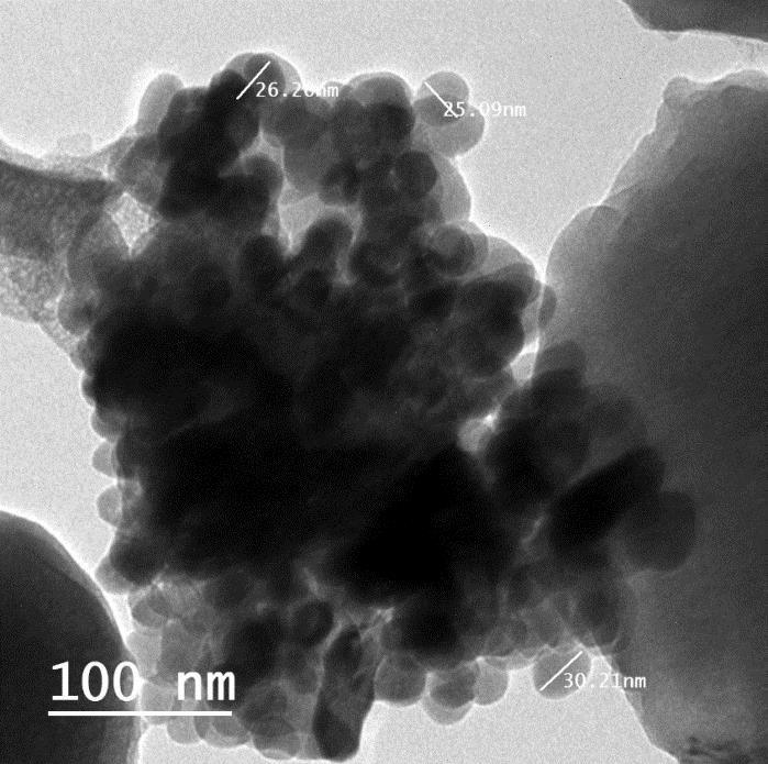
ABSTRACT: Antibiotic-resistant bacteria have become one of the major issues and concerns worldwide. For the past years, scientists have investigated the use of treatments in the nano-scale. Nanomaterials, such as metal oxide nanoparticles, have shown promising results due to their antibacterial properties. The aim of this study was to investigate the efficiency of in vitro antibacterial activity of zinc oxide nanoparticles (ZnO NPs) alone and in combination with different antibiotics against avian pathogenic Escherichia coli. In this study, ZnO NPs were synthesized using direct precipitation method. Physical characteristics of ZnO NPs were confirmed using X-ray diffraction and transmission electron microscopy. Antibacterial resistance pattern of 10 antibiotics including amoxicillin, ciprofloxacin, enrofloxacin, gentamicin, doxycycline, levofloxacin, trimethoprim/sulfamethoxazole, tetracycline, spiramycin, and streptomycin, in addition to different concentrations of ZnO NPs, was determined by disc diffusion method on 10 avian pathogenic E. coli (APEC). The results showed that 50% of the strains were resistant to all antibiotics, while the rest were found to be sensitive to one or two antibiotics. The best concentration of ZnO NPs was 50 mg/disk, which showed greater zones than that of other used concentrations (25, 12.5, 6.25, 3.125, and 1.56 mg/disk). The combination of spiramycin and gentamycin with ZnO NPs showed a synergistic effect while the combination of ZnO NPs with ciprofloxacin, enrofloxacin, and streptomycin showed an antagonistic effect. No antibacterial effect was observed in combination of ZnO NPs with other used antibiotics. This study recommends in vivo evaluations to confirm the results.
Keywords: Antibiotic, Escherichia coli, Nanoparticle, Zinc Oxide
[Full text-PDF] [XML] [HTML] [ePub] [Crossref Metadata] [Google Scholar]
Immunological Study on Salmonellae Isolated from Different Sources
Shedeed EA, El-Hariri MD, Nasef SA and El Jakee J.
J. World Poult. Res. 10(2S): 285-291, 2020; pii: S2322455X2000034-10; DOI: https://dx.doi.org/10.36380/jwpr.2020.34
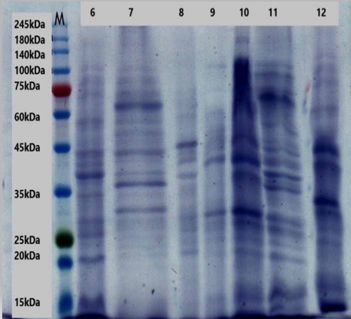
ABSTRACT: Salmonella infection is a critical veterinary and medical problem worldwide and is a major issue in the food industry. Non-typhoidal Salmonella is known as an important pathogen causing gastroenteritis. The Outer Membrane Proteins (OMPs) of Gram negative bacteria are significant for virulence, host immune responses and drug therapy targets. Enhanced diagnosis of live poultry colonized with Salmonella species is required to avoid foodborne diseases. The present study was based on molecular characterization of OMPs among four Salmonella serovars (S. Typhimurium, S. Enteritidis, S. Kentucky and S. Anatum) using sodium dodecyl sulfate-polyacrylamide gel electrophoresis. The OMPs profiling showed more than 70 protein bands ranged in size from 208 kDa to below 16 kDa which were detected using Total Lab 1D 12.2 software. All Salmonella strains had a band at 54-60 kDa, 45-53 kDa, 36-39 kDa and 26-31 kDa. Eleven strains exhibited a band at 41-46 kDa and 33-35 kDa. Nine strains had a band at 61-69 kDa. Eight strains exhibited a band at 135-145 kDa and 72-79 kDa. Seven strains had a band at 108-123 kDa and 83-91 kDa. In the Western blot analysis, the prepared hyperimmune anti serum of each Salmonella serovars reacted with the 35 kDa protein band. It is concluded that the identification of novel immunogenic proteins would be useful in developing ELISA-based diagnostic assays with a higher specificity.
Key words: Outer Membrane Proteins, Salmonella, SDS-PAGE, Western blotting
[Full text-PDF] [XML] [HTML] [ePub] [Crossref Metadata] [Google Scholar]
Detection of Virulence Genes in Bacillus cereus isolated from Meat Products Using PCR
Abd El Tawab AA, El-Hofy FI, Abou El Roos NA and El-morsy DA.
J. World Poult. Res. 10(2S): 292-298, 2020; pii: S2322455X2000035-10; DOI: https://dx.doi.org/10.36380/jwpr.2020.35
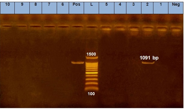
ABSTRACT: Bacillus cereus is an opportunistic pathogen that can cause food poisoning in humans as a result of consuming foods containing toxins or bacteria. In this study, the incidence of B. cereus and its virulence genes in meat products was investigated. Isolation of B. cereus was performed using selective PEMBA media and confirmed by morphological and biochemical tests and Vitek2 compact system. The incidence of B. cereus strains in beef and chicken meat products was 28%. The incidence of Bacillus cereus in frozen rice kofta, frozen kobiba-shami, chicken pane, and chicken nuggets was 16%, 24%, 28%, and 44%, respectively. Moreover, the result of multiplex PCR of virulence genes of groEL gene (533bp), Hbl gene (1091 bp), Nhe gene (766 bp) and Cytk gene (421bp) indicated that groEl gene, Nhe gene, Cytck gene was found in 100% of B. cereus isolated from different meat products, while Hbl gene was detected in 10% of isolates. The results demonstrate that meat products represent a threat to public health through the transmission of Bacillus cereus.
Key words: Bacillus cereus, Beef meat, Chicken meat, PCR, Virulence genes, VITEK2
[Full text-PDF] [XML] [HTML] [ePub] [Crossref Metadata] [Google Scholar]
Development of a Duplex Real-time PCR for Differentiation of Salmonella Typhimurium and Monophasic Serovars
Abd El-Lattief A, Marouf Sh, El-Bialy A and El-Jakee J.
J. World Poult. Res. 10(2S): 299-325, 2020; pii: S2322455X2000036-10; DOI: https://dx.doi.org/10.36380/jwpr.2020.36
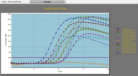
ABSTRACT: Salmonella Typhimurium is the most Salmonella serovar causing acute gastroenteritis and diarrhea. Serovar 1, 4, [5], 12: i:- is considered a monophasic variant of S. Typhimurium that threaten public health. Fifty-eight serologically confirmed Salmonella strains were investigated by PCR using 16S rRNA and fliC genes. All 58 strains harbored 16S rRNA while 21 strains harbored fliC gene that included S. Typhimurium (12), S. Kentucky (6), Salmonella variant strain serotype 1, 4, [5],12:i:- (1), S. Lagos (1), and S. Kedougou (1). A duplex TaqMan real-time PCR was performed for differentiating between biphasic S. Typhimurium and monophasic serovar 1, 4, [5], 12:i:- using fljB1, 2 and fliB/IS200 in the fliA-fliB intergenic region. Ten out of twelve S. Typhimurium harbored fljB 1, 2, while Salmonella variant strain serotype 1, 4, [5], 12:i:- lacked this gene. Thirteen strains (12 S. Typhimurium and the variant strain serotype 1, 4, [5], 12:i:-) were positive for fliB/IS200 that is a specific gene for S. Typhimurium (biphasic and monophasic ). The result of duplex TaqMan real-time PCR indicated that 10 S. Typhimurium strains were biphasic while two S. Typhimurium strains and the variant strain serotype 1, 4, [5], 12:i:- lack fljB1,2 and had fliB/IS200 were monophasic S. Typhimurium. It is noticed that prolonged subculture and repeat phase inversion method leads to the formation of flakes that in turn cause wrongly serotype identification, therefore, real-time PCR is rapid and can be used for identifying and differentiating between biphasic and monophasic S. Typhimurium.
Key words: Biphasic and monophasic S. Typhimurium, flj gene, Real-time PCR, Salmonella.
[Full text-PDF] [XML] [HTML] [ePub] [Crossref Metadata] [Google Scholar]
Previous issue | Next issue | Archive
Poultry Research Center Annex  |
International Conference of The Egyptian Poultry Forum (ICEPF 2020 February)
ICEPF 2020 has been held 1st of March 2020 in Hurghada, Egypt, by the Egyptian Poultry Forum Foundation as authorized partner for the SCIENCELINE International journals (WVJ, JWPR, OJAFR) representing Egypt and MENA region.
List of editors and reviewers
Ahmed A. Ali, MVSc, PhD, IFBA Certified Professional, Lecturer of Poultry Diseases; Poultry Diseases Department, Faculty of Veterinary Medicine, Beni-suef University, Beni-Suef 62511, EGYPT, Email: ahmed.ali1@vet.bsu.edu.eg
Ahmed Abdel-Kareem Abuoghaba, M.Sc., PhD, Dept. of poultry Production, Faculty of Agriculture, Sohag University, Sohag, EGYPT
Ahmed Ragab Elbestawy, PhD, Assistant Lecturer of poultry diseases, Faculty of Veterinary Medicine- Damanhour University, EGYPT
Ali Othman Othman Al-Ghamdi, Professor of Parasitology, Baha University, Saudia Arabia.
Amine Berghiche; Teacher-researcher in fields of Veterinary Biostatistics, Antibiotics, Meat quality, Broiler); PhD of Agronomy, Souk Ahras University; ALGERIA; Email: amine_berghiche@yahoo.com
Arman Moshaveri, DVM, Faculty of Veterinary Medicine, Karaj Branch, Islamic Azad University, Karaj, IRAN
Avinash Warundeo Lakkawar, MVSc, PhD, Associate Professor, Department of Pathology, Rajiv Gandhi Institute of Veterinary Education and Research (RIVER), Kurumbapet, Pondicherry- 605009, INDIA
Carlos Daniel Gornatti Churria, Med. Vet., Dr. Cs. Vet., Lecturer; Cátedra de Patología de Aves y Pilíferos, Facultad de Ciencias Veterinarias, Calle 60 y 118 s/n, Universidad Nacional de La Plata, Pcia. Bs. As., ARGENTINA
Daryoush Babazadeh, DVM, DVSc, PhD of Avian/Poultry Diseases, School of Veterinary Medicine, Shiraz University, Shiraz, IRAN
Eilyad Issabeagloo, PhD, Assistant Prof. of Pharmacology; Dep. Basic Sciences, Faculty of medical Sciences, Tabriz Branch, Islamic Azad University, Tabriz, IRAN
Farooz Ahmad Lone, PhD, Assistant Prof. Semen Cryopreservation, Estrous induction, In vitro maturation and fertilization, Reproductive diseases; Division of Animal Reproduction, Gynecology and Obstetrics, Faculty of Veterinary sciences and animal husbandry, Shere-Kashmir University of agricultural sciences and technology of Kashmir, 190006, J&K, INDIA
Ghulam Abbas Muhammad Jameel, PhD, Poultry Science, Animal Sciences Institute, University of Agriculture Faisalabad, PAKISTAN
Hadi Haghbin Nazarpak, PhD. Poultry Diseases, Department of clinical sciences, Faculty of Veterinary Medicine, Garmsar Branch, Islamic Azad University, Garmsar, IRAN.
Hazim Jabbar Al-Daraji, PhD, Prof. of Avian Reproduction and Physiology; College of Agriculture, University of Baghdad, IRAQ
Huthail Najib Abdulrahman, Professor of Poultry Nutrition, King Faisal University, Saudia Arabia.
Ir. Irfan H. Djunaidi, Dr. Poultry Nutrition, Animal Science Faculty. Universitas Brawijaya, Indonesia.
Samera Lateef Hameed Al-Ameen, President of iraqi veterinary medical syndicate, Iraq.
John Cassius Moreki, PhD, Nutrition - Poultry Science, Breeders; Department of Animal Science and Production, Botswana College of Agriculture, Gaborone, BOTSWANA
Karamala Sujatha, MVSc, PhD, Associate Professor, Department of Veterinary Pathology, College of Veterinary Science, Sri Venkateswara Veterinary University, Tirupati – 517502, Andhra Pradesh, INDIA
Karim Mohamed El-Sabrout; PhD, Assistant Prof., University of Alexandria, Faculty of Agriculture, Department of Poultry Production, Alexandria, EGYPT
Khenenou Tarek; PhD of Avian Diseases, Histopathology; Institut des sciences vétérinaires et agronomiques. Département vétérinaire, Université, Mohamed Chérif Messaadia de Souk-Ahras, ALGERIA; Email: tarekkheneneou @yahoo.fr
Konstantinos Koutoulis; DVM, PhD; Avian Pathology, University of Thessaly, Terma Trikalon 224, 43100 Karditsa, GREECE
Maha Mohamed Hady Ali, PhD, Professor of Nutrition and clinical Nutrition, Cairo University, EGYPT
Mahmoud El-Said sedeik, PhD, Associate Professor of Poultry diseases; Department of Poultry and fish Diseases, Faculty of Veterinary Medicine, Alexandria University, EGYPT
Maryam Karimi Dehkordi, PhD, Veterinary Clinical Pathology, Department of clinical Sciences, Faculty of Veterinary Medicine, Shahrekord Branch, Islamic Azad University, Shahrekord, Iran. E.mail: ma_karimivet58@yahoo.com
Mohamed Shakal, Professor & Head of Poultry Diseases Department, Faculty of Veterinary Medicine, Cairo University, EGYPT; Director of the Endemic and Emerging Poultry Diseases Research Center, Cairo University, Shek Zaed Branch, EGYPT; Chairman of The Egyptian Poultry Forum Scientific Society. REPRESENTATIVE FOR EGYPT & MENA REGION. Email: shakal2000@gmail.com
Mohammad A. Hossain, PhD, Associate Professor, Department of Dairy and Poultry Science, Chittagong Veterinary and Animal Sciences University; Khulshi; Chittagong; Bangladesh
Mohammed Muayad Taha, Associate Prof., PhD of Animal physiology, University Pendidikan Sultan Idris, Malaysia 2017. ORCID: 0000-0002-8106-6460
Moharram Fouad El-Bassiony, Associate Professor of Animal Physiology, Animal and Poultry Physiology Department, Desert Research Center, www.drc.gov.eg; PhD, Faculty of Agriculture, Cairo Univ., Cairo, EGYPT
Muhammad Moin Ansari, BVSc & AH, MVSc, PhD (IVRI), NET (ICAR), Dip.MLT, CertAW, LMIVA, LMISVS, LMISVM, MHM, Sher-e-Kashmir University of Agricultural Sciences and Technology of Kashmir, Faculty of Veterinary Sciences and Animal Husbandry, Division of Veterinary Surgery and Radiology, Shuhama, Alastang, Srinagar-190006 Jammu & Kashmir, INDIA
Muhammad Saeed, PhD candidate, Animal Nutrition and Feed Science, College of Animal Sciences and Feed technology, Northwest A&F University, Yangling, 712100, CHINA
Neveen El Said Reda El Bakary, Ph.D., Assistant Prof. of Comparative anatomy, Ultrastructure, Histochemistry, Histology; Department of Zoology, Faculty of Science, Mansoura University, New Damietta, EGYPT
Reihane Raeisnia, DVM, School of Veterinary Medicine, Ferdowsi University of Mashhad, Mashhad, Iran; Email: rhn.raeisnia@gmail.com
Roula Shaaban Ibrahim Hassan, Dr., President of Emirates Veterinary Association, UAE
Saeid Chekani Azar, PhD, DVM, Animal Physiology; Faculty of Veterinary Medicine, Atatürk University, TURKEY
Salah M. Hassan, Prof. Dr., College of Veterinay Medicine, Al-Qasim green University, Iraq.
Salwan Mahmood Abdulateef, PhD, Assistant Lecturer - Behavior & Environmental Physiology of Poultry; College Of Agriculture, University of AL-Anbar, Republic of IRAQ
Sami Abd El-Hay Farrag, PhD, Poultry Production Dep., Faculty of Agriculture, Menoufia University, Shebin El-Kom, Menoufia, EGYPT
Sandeep Kumar Sharma, PhD, Assistant professor & In-charge; Department of Veterinary Microbiology and Biotechnology; Post Graduate Institute of Veterinary Education and Research; Rajasthan University of Veterinary and Animal Sciences, Jamdoli, Jaipur-302031, INDIA; Email: drsharmask01@hotmail.com
Shahid Nazir, Avian Pathology; School of Veterinary Medicine, Wollo University, Dessie, Amhara Region, ETHIOPIA
Shahriar Behboudi, Prof. Dr., Head of Avian Immunology, Pirbright Institute and Surrey University, UK.
Shahrzad Farahbodfard, DVM, School of Veterinary Medicine, Ferdowsi University of Mashhad, Mashhad, Iran; Email: shahrzad.vetmed@gmail.com
Siti Azizah, Associate Professor of Animal Science Faculty, Universitas Brawijaya, Indonesia.
Thakur Krishna Shankar Rao, PhD, Assistant professor, Vanabandhu College of Veterinary Science & Animal Husbandry, Navsari Agricultural University, Navsari Gujarat, INDIA
Thandavan Arthanari Kannan, PhD, Full professor, Centre for Stem Cell Research and Regenerative Medicine Madras Veterinary College Tamil Nadu Veterinary and Animal Sciences university Chennai-600007, INDIA
Tugay AYAŞAN, PhD, Cukurova Agricultural Research Institute, PK: 01321, ADANA, TURKEY
Wafaa Abd El-Ghany Abd El-Ghany, PhD, Associate Professor of Poultry and Rabbit Diseases; Department of Poultry Diseases, Faculty of Veterinary Medicine, Cairo University, Giza, EGYPT
Wesley Lyeverton Correia Ribeiro, MSc, DVM, Animal Health, Veterinary Parasitology, and Public Health, Animal welfare and Behavior; College of Veterinary Medicine, State University of Ceará, Av. Paranjana, 1700, Fortaleza, BRAZIL
![]() This work is licensed under a Creative Commons Attribution 4.0 International License (CC BY 4.0).
This work is licensed under a Creative Commons Attribution 4.0 International License (CC BY 4.0).


